How do we see and learn? (Part 3: Memory)


Everyone wants to learn faster and effectively.

One way is to understand how the learner’s brain processes in the learning environment. Learning is the process of taking new information in your working memory and integrating it with existing knowledge in your long-term memory.  Once it’s in long-term memory you can recall it and transfer the knowledge to the real world.


  • Working memory:  Your working memory is good at processing information, but it can only hold so much at one time.  All of your active thinking happens in the working memory.
  • Long-term memory:  Your long-term memory is your storage center and holds your existing knowledge.  In the learning process, you are connecting the new information to prior knowledge.  As you actively process information, you are swapping it between working and long-term memory.

The working memory is like a white board where you can do a lot of calculations and diagramming on the fly.  On the white board, you need space to both write down information (temporary storage) and do your problem-solving (active processing).

Often the problem is that you only have so much space.  As the white board gets cluttered with information, you run out of room to work.  That means you need to record the important information and free up space to do more work on the white board.

One way to capture the information is to create post-it notes (long-term memory) to record the information on the white board.  Once you you have the notes, you are free to erase the white board and do more work.  And, if you needed to recall what you did earlier, all you have to do is look at one of your notes.


As you go through an learning process, what you see and hear enters your working memory where it is temporarily stored.  Your brain actively processes the new information and integrates it with what you have stored in your long-term memory.

So, your brain is doing these things:

  1. Receiving new information
  2. Actively processing the information
  3. Integrating the information with long-term memory

(ref: http://www.articulate.com/rapid-elearning/2007/10/)

There are numerous ways been suggested by the researchers about effective ways to transfer information to the learners. These are used to promote clear processing and life long memory of the information and teachers in classroom happen to use coloured movie clips and pens. Most of the schools have recognized the importance in providing resources and been proved to be effective.
How do we see? (Part2: Brain and vision)

Colour blindness and night blindness

Most forms of colour blindness, an inherited inability to distinguish between certain colours, result from the absence or a deficiency of one of the three cone photopigments. The most common type is red-green colour blindness, in which a photopigment sensitive to orange-red light or green light is missing. As a result, the person cannot distinguish between red and green. Prolonged vitamin A deficiency and the consequent below normal amount of rhodopsin may cause night blindness – an inability to see well at low light levels.

…………………………………………………………………

The visual pathway

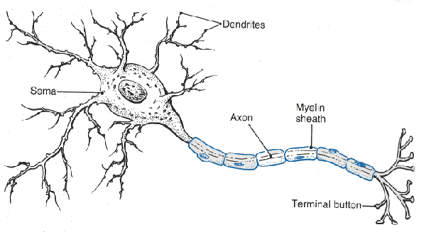
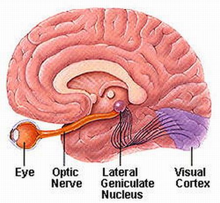
After considerable processing of visual signals in the retina at synapses among the various types of neurons, the axons of retinal ganglion cell provide output from the retina to the brain.

They exit the eyeball as the optic nerve (cranial nerve II).

(http://www.msstrength.com/ms-optic-nerve-attacks-and-symptoms/)

…………………………………………………………………

Process of Visual Input in the Retina

Within the retina, certain features of visual input are enhanced while other features may be discarded. This is because there are only 1million ganglion cells but 126 million photoreceptors.

Once receptors in rods and cones receives message, the message is spread through the synaptic terminals of vision neurons. Everything that can be seen by one eye is that eye’s visual field. Because our eyes are located anteriorly in the head, the visual field of the two eyes overlap considerably. We have binocular vision due to the large region where the visual fields of the two eyes overlap. The visual field of each eye is divided into two regions. Moreover, visual information from the right half of each visual field is conveyed to the left side of the brain, where as visual information from the left half of each visual field is conveyed to the right side of the brain.

(ref: http://www.arn.org/docs/glicksman/120104%20fig3.jpg)

  1. Axons of all retinal ganglion cells in one eye exit the eyeball at the optic disc and form the optic nerve on that side.
  2. At the optic chiasm, axons from the temporal half of each retinal do not cross but continue directly to the thalamus on the same side.
  3. In contrast, axons from the nasal half of each retina cross and continue to the opposite thalamus.
  4. Axon branches of the retinal ganglion cells project to the mid brain, where they participate in circuits that govern constrictions of pupils in response to light and co-ordination of head and eye movements. Hypothalamus also establishes the patterns of sleep and other activities that occur on a daily schedule in response to intervals of light and darkness.
  5. The axons of thalamic neurons form the optic radiation as they project from the thalamus to the primary visual area of the cortex on the same side.

(ref: http://www.thebrainwiki.com/uploads/Forebrain/thalamus.jpg)


…………………………………………………………………


Being a part of the cerebrum, They are located at the edges of the brain. Each temporal lobe deals with auditory processing and semantics of speech and vision. The temporal lobe hosts the hippocampus and is therefore involved in the formation of memories. Their function include Emotional Responses, Hearing, Memory and Speech.


How do we see? (Part1: The Eye)

In some ways the eye is like a camera: Its optical elements focus an image of some object on a light-sensitive “film – the retina – while ensuring the correct amount of light to make the proper “exposure”.

……………………………………………………………………………………………

When your eyelids are open, light enters your eye through a circular hole called the pupil and is focussed by a lens onto the light sensitive retina attached to the back of the eye.

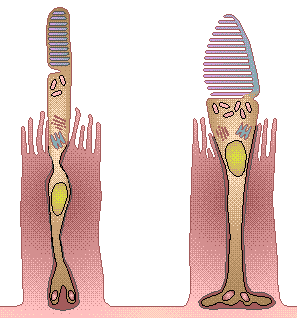
The size of the pupil can be adjusted to allow more light to enter when the environment is dim, and less light when it’s bright.   There are about 126 million sensory cells in the retina, both cone-shaped cells which are color-sensitive and rod-shaped cells which aren’t color-sensitive but can detect low levels of light, useful for night vision.

Most cameras work in the same way as the eye – when the shutter is open, light enters a roughly circular hole called theaperture and is focussed by a lens onto a light sensitive medium at the back of the camera, either film or an electronic sensor.   Some types of camera, like a pinhole camera, don’t have a lens, and some digital cameras don’t have a shutter; nevertheless, understanding how these things work will help make your photographs better. (ref: Flying Kiwi)


……………………………………………………………………………………………

To understand how the eye forms clear images of objects on the retina, we must examine three processes:

  1. The refraction (bending) of light by the lens and cornea
  2. The change in shape of the lens
  3. Narrowing of the pupil

More information about vision – http://www.accessexcellence.org/AE/AEC/CC/vision_background.php

……………………………………………………………………………………………

Brains learns to see

When light rays traveling through a transparent substance pass into a second transparent substance with a different density, they bend at the junction between the two. This bending is called refraction. As light rays enter the eye, they are refracted at the anterior and posterior surface of the cornea. Both surfaces of the lens of the eye further refract the light rays so they come into exact focus on the retina. Images focused on the retina are inverted; they are upside down. The reason the world does not look inverted and reversed is that the brain “learns” early in life to co-ordinate visual images with the orientations of objects. The brain stores the inverted and reversed images we acquired when we first reached for and touched objects and interprets those visual images as being correctly oriented in space.

……………………………………………………………………………………………

Physiology of Vision: Photoreceptors and photopigments

The first step in visual transduction is absorption of light by a photopigment, a colored protein that undergoes structural changes when it absorbs light. The single type of photopigment in rods is rhodopsin. Three different cone photopigments are present in the retina, one in each of three types of cones. Colour vision results from different colors of light selectively activating the different cone photopigments.  All photopigments associated with vision contain two parts: retinal and opsin. Different opsins permit the rods and cones to absorb different colours (wavelength) of incoming light. Rhodopsins absorbe blue to green light(colour) most effectively, where as the three different cone photopigments most effectively absorb blue, green, or yellow-orange light(and colour).