
In some ways the eye is like a camera: Its optical elements focus an image of some object on a light-sensitive “film – the retina – while ensuring the correct amount of light to make the proper “exposure”.
……………………………………………………………………………………………
When your eyelids are open, light enters your eye through a circular hole called the pupil and is focussed by a lens onto the light sensitive retina attached to the back of the eye.
The size of the pupil can be adjusted to allow more light to enter when the environment is dim, and less light when it’s bright. There are about 126 million sensory cells in the retina, both cone-shaped cells which are color-sensitive and rod-shaped cells which aren’t color-sensitive but can detect low levels of light, useful for night vision.
Most cameras work in the same way as the eye – when the shutter is open, light enters a roughly circular hole called theaperture and is focussed by a lens onto a light sensitive medium at the back of the camera, either film or an electronic sensor. Some types of camera, like a pinhole camera, don’t have a lens, and some digital cameras don’t have a shutter; nevertheless, understanding how these things work will help make your photographs better. (ref: Flying Kiwi)

- To learn the structures of an eye – http://www.preventblindness.org/vlc/how_we_see.htm
- Read how camera works – http://electronics.howstuffworks.com/camera.htm
……………………………………………………………………………………………
To understand how the eye forms clear images of objects on the retina, we must examine three processes:
- The refraction (bending) of light by the lens and cornea
- The change in shape of the lens
- Narrowing of the pupil

More information about vision – http://www.accessexcellence.org/AE/AEC/CC/vision_background.php
……………………………………………………………………………………………
Brains learns to see
When light rays traveling through a transparent substance pass into a second transparent substance with a different density, they bend at the junction between the two. This bending is called refraction. As light rays enter the eye, they are refracted at the anterior and posterior surface of the cornea. Both surfaces of the lens of the eye further refract the light rays so they come into exact focus on the retina. Images focused on the retina are inverted; they are upside down. The reason the world does not look inverted and reversed is that the brain “learns” early in life to co-ordinate visual images with the orientations of objects. The brain stores the inverted and reversed images we acquired when we first reached for and touched objects and interprets those visual images as being correctly oriented in space.
……………………………………………………………………………………………
Physiology of Vision: Photoreceptors and photopigments
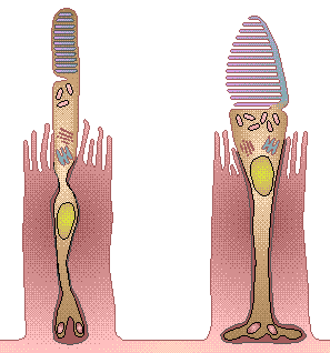
The first step in visual transduction is absorption of light by a photopigment, a colored protein that undergoes structural changes when it absorbs light. The single type of photopigment in rods is rhodopsin. Three different cone photopigments are present in the retina, one in each of three types of cones. Colour vision results from different colors of light selectively activating the different cone photopigments. All photopigments associated with vision contain two parts: retinal and opsin. Different opsins permit the rods and cones to absorb different colours (wavelength) of incoming light. Rhodopsins absorbe blue to green light(colour) most effectively, where as the three different cone photopigments most effectively absorb blue, green, or yellow-orange light(and colour).