Colour blindness and night blindness
Most forms of colour blindness, an inherited inability to distinguish between certain colours, result from the absence or a deficiency of one of the three cone photopigments. The most common type is red-green colour blindness, in which a photopigment sensitive to orange-red light or green light is missing. As a result, the person cannot distinguish between red and green. Prolonged vitamin A deficiency and the consequent below normal amount of rhodopsin may cause night blindness – an inability to see well at low light levels.
…………………………………………………………………
The visual pathway
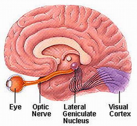
After considerable processing of visual signals in the retina at synapses among the various types of neurons, the axons of retinal ganglion cell provide output from the retina to the brain.
They exit the eyeball as the optic nerve (cranial nerve II).
(http://www.msstrength.com/ms-optic-nerve-attacks-and-symptoms/)
…………………………………………………………………
Process of Visual Input in the Retina
Within the retina, certain features of visual input are enhanced while other features may be discarded. This is because there are only 1million ganglion cells but 126 million photoreceptors.
Once receptors in rods and cones receives message, the message is spread through the synaptic terminals of vision neurons. Everything that can be seen by one eye is that eye’s visual field. Because our eyes are located anteriorly in the head, the visual field of the two eyes overlap considerably. We have binocular vision due to the large region where the visual fields of the two eyes overlap. The visual field of each eye is divided into two regions. Moreover, visual information from the right half of each visual field is conveyed to the left side of the brain, where as visual information from the left half of each visual field is conveyed to the right side of the brain.

(ref: http://www.arn.org/docs/glicksman/120104%20fig3.jpg)
- Axons of all retinal ganglion cells in one eye exit the eyeball at the optic disc and form the optic nerve on that side.
- At the optic chiasm, axons from the temporal half of each retinal do not cross but continue directly to the thalamus on the same side.
- In contrast, axons from the nasal half of each retina cross and continue to the opposite thalamus.
- Axon branches of the retinal ganglion cells project to the mid brain, where they participate in circuits that govern constrictions of pupils in response to light and co-ordination of head and eye movements. Hypothalamus also establishes the patterns of sleep and other activities that occur on a daily schedule in response to intervals of light and darkness.
- The axons of thalamic neurons form the optic radiation as they project from the thalamus to the primary visual area of the cortex on the same side.

(ref: http://www.thebrainwiki.com/uploads/Forebrain/thalamus.jpg)
…………………………………………………………………

Being a part of the cerebrum, They are located at the edges of the brain. Each temporal lobe deals with auditory processing and semantics of speech and vision. The temporal lobe hosts the hippocampus and is therefore involved in the formation of memories. Their function include Emotional Responses, Hearing, Memory and Speech.